CLOSE

Date:
2009
Journal:
22nd European Conference on Biomaterials ESB 2009 - 07-11th September, Lausanne (Switzerland)
Author:
G. Pertici, G. Perale, S. Maccagnan, M. Müller
Link:

INTRODUCTION
Scaffolds for bone tissue engineering (t.e.) should ensure both mechanical stability and adequate strength and show a completely interconnected porous network for cell migration and proliferation, while also providing specific signals for bone regeneration. These issues are very well know ever since early maxillo-facial surgery in the 80’s. Moreover, connective tissue adhesion should be promoted. Biopolymers, bioglass and bioceramics are often considered to satisfy these requirements at least in part. Here, Industrie Biomediche Insubri S/A developed and produced a new composite 3D scaffold thought for bone t.e., particularly for maxillo-facial surgery, using a novel concept based on biomaterial assembly, thus avoiding drawbacks of interfacial bonding.
MATERIALS AND METHODS
This novel matrix has a composite structure based on a bone graft specifically reinforced with biodegradable polymers and bioactive agents. The bone grafts allow to maintain the adequate 3D-structure and porosity, the biopolymers permit to achieve good mechanical properties while bioactive agents promote cell adhesion and proliferation. All used components are approved for human use and scaffolds are produced according to GMP standards at the Micro-Sphere S/A facility. Both process and product are proprietary.
Microstructure was evaluated by SEM imaging (Evo 50EP, Zeiss-Cambridge Instruments, Germany). Handling tests were performed by surgeons to assess applicability in oral surgery. Preliminary in vitro tests with bovine chondrocytes were performed to evaluate the ability to support and promote cells proliferation within the matrix.
RESULTS
This novel composite 3D scaffold resulted feasible for oral surgery, being both easy to shape and resistant to screws and fixation manoeuvres. Moreover, SEM imaging confirmed that its microscopic structure resembles that of human cortical bone. Cell proliferation, assessed by Alamar Blue™ test, and viability resulted both positive and fully comparable in terms of values referred to controls. Further confirmations came from morphological analysis performed via SEM imaging.
CONCLUSIONS
Data collected represent a positive proof of concept for the developed process and the application of this new type of matrix as scaffold for bone tissue regeneration. Morphological analysis of structure confirmed the presence of a well diffused cortical bone human-like porosity. Preliminary mechanical investigation showed easy shaping by common surgical instruments in order to replicate and thus replace bone defects. Furthermore, preliminary biological investigations showed scaffolds to be promising substrates for cell adhesion and growth.
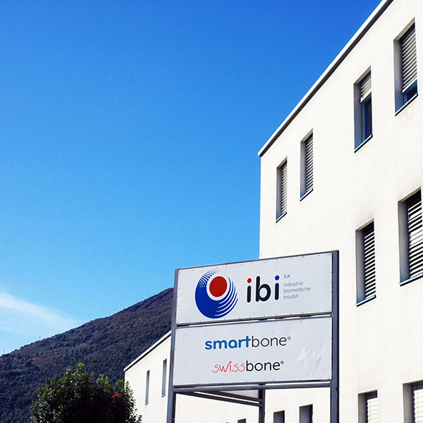
IBI SA
Industrie Biomediche Insubri SA
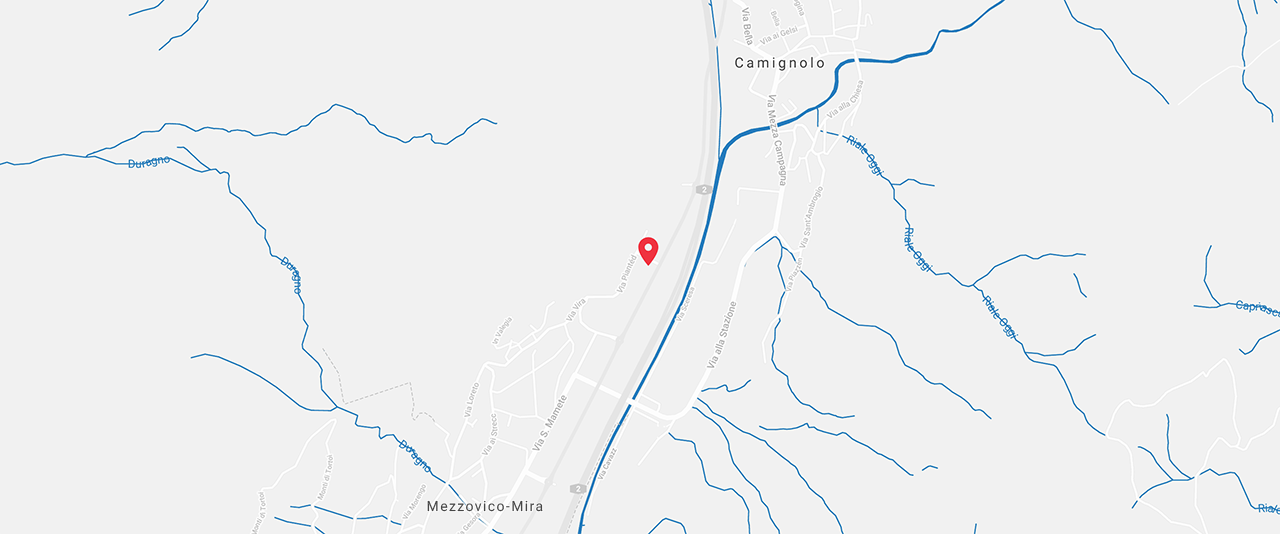
via Cantonale 67, CH-6805 Mezzovico-Vira, Switzerland
t. +41 91 93.06.640
f. +41 91 220.70.00